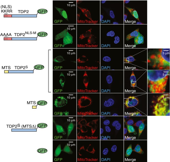
Figure 3. TDP2S bears a mitochondrial targeting sequence.

Fluorescence microscopy images of HCT116 cells transfected with the indicated TDP2 constructs tagged with C‐terminal GFP (schemes at left of each panel). Columns from left to right show GFP (green), MitoTracker® Deep Red (red), DAPI (blue) and merged images with selective zoomed‐in fields. NLS is the nuclear localization sequence, NLS‐M is the nuclear localization sequence‐mutated, MTS is the mitochondrial targeting sequence, and TDP2S (MTSΔ) is TDP2S lacking the N‐terminal MTS.
