-
A
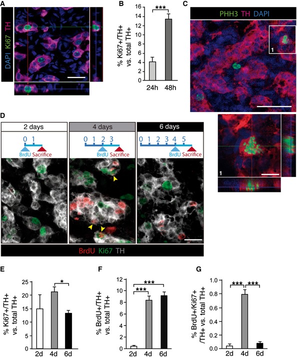
Immunohistochemical analysis of carotid body (CB) removed from rats subjected to hypoxia for 24 h. Image shows tyrosine hydroxylase (TH; red)‐positive cells expressing the cell cycle marker Ki67 (green).
-
B
Quantification of the frequency of proliferating CB TH+ cells after 24 and 48 h in hypoxia (n = 4–6 carotid bodies per time point).
-
C
Confocal micrograph showing an example of a CB TH+ cell (red) in M phase of the cell cycle, thus accounting for its positivity to phosphorylated histone H3 (PHH3; green).
-
D
Time course of CB TH+ (gray) cell proliferation, studied by Ki67 staining and BrdU incorporation during the 48 h prior sacrifice (see diagrams on top). Yellow arrowheads point to TH/Ki67/BrdU triple‐positive cells.
-
E–G
Quantifications of the time course experiment shown in (D), highlighting the transient nature of adult CB TH+ cell proliferation in response to chronic hypoxia (n = 3–6 carotid bodies per time point).
Data information: Scale bars: 20 μm in (A), 40 μm in (C), 10 μm in (C1), and 15 μm in (D). Data in bar graphs are presented as mean ± SEM. *
P < 0.05, ***
P < 0.001 (Student's
t‐test).
Source data are available online for this figure.

