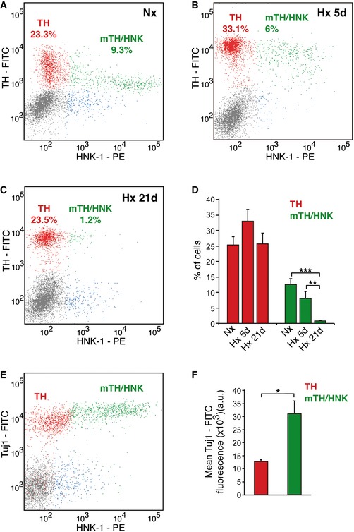
Figure 3. mTH+/HNK+ cells are immature CB neuroblasts.

-
A–CFlow cytometric analysis of dispersed CB cells from normoxic (Nx), 5d hypoxic (Hx), or 21d hypoxic (Hx) rats, stained for TH and HNK‐1. Note how mTH+/HNK+ cells convert into TH+/HNK− mature glomus cells upon exposure to hypoxia.
-
DQuantification of the flow cytometry analysis shown in (A–C) (n = 5–7 rats per group).
-
EFlow cytometry analysis of Tuj1 expression in dispersed CB cells from normoxic animals. Note how mTH+/HNK+ cells express higher levels of Tuj1 than mature TH+/HNK− cells, confirming their immature phenotype.
-
FQuantification of the mean Tuj1 expression in TH+/HNK− mature glomus cells versus mTH+/HNK+ immature neuroblasts (n = 3 independent experiments with a total of 10 rats).
