
Figure 2.
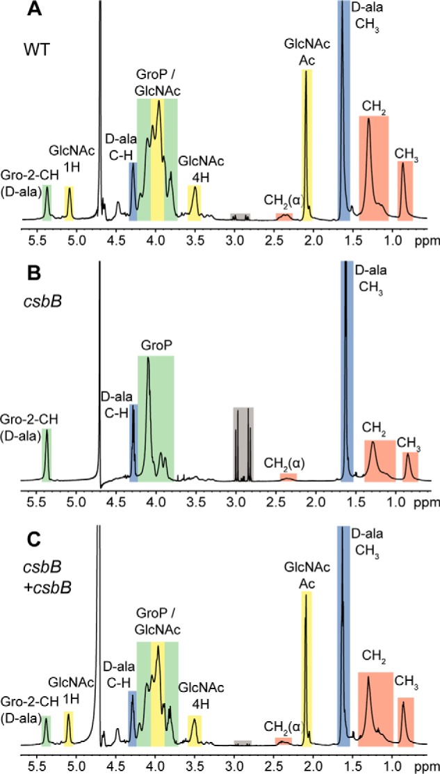
NMR analysis of LTA isolated from WT B. subtilis 168, csbB mutant, and complementation strains. Shown are NMR spectra of LTA derived from B. subtilis strains 168 (WT) (A), csbB mutant (B), and the csbB+csbB complementation strain (C). Colored boxes and labels indicate nonexchangeable protons derived from the different LTA components. Peaks were assigned as described previously (17, 37–39). The different peaks for the protons and acetyl group of GlcNAc are labeled with 1H, 4H, and Ac, respectively. Gray boxes highlight peaks resulting from residual citrate, a buffer component used during the LTA purification procedure. The spectra are representative of three independent experiments.