
Figure 4.
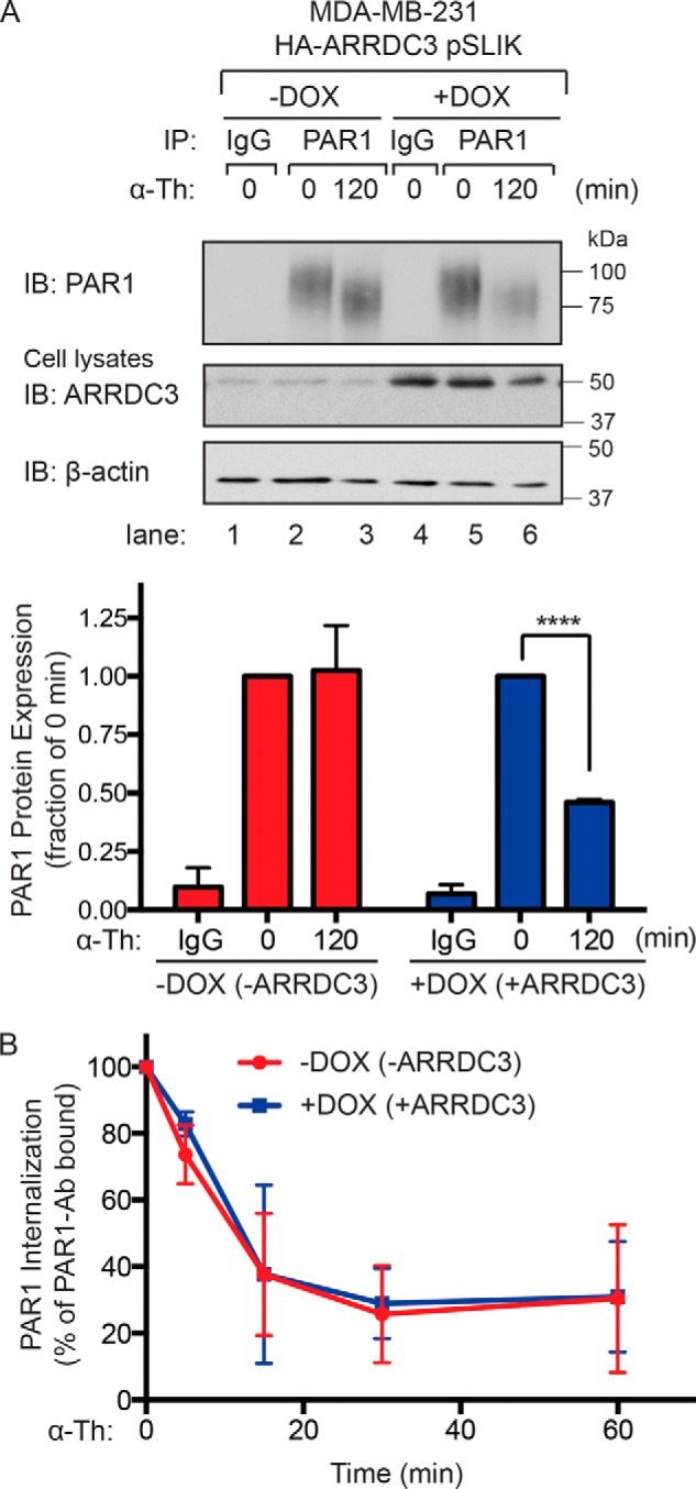
Thrombin-induced PAR1 lysosomal degradation is restored in cells re-expressing ARRDC3. A, MDA-MB-231 HA-ARRDC3 pSLIK cells incubated with or without 1 μg/ml DOX for 48 h were treated with 10 nm α-thrombin for the indicated times, lysed, and immunoprecipitated with anti-PAR1 WEDE antibody or anti-IgG control. Cell lysates were immunoblotted for HA-ARRDC3 and β-actin expression. The data (mean ± S.D. (error bars), n = 3) are represented as the fraction of PAR1 protein remaining relative to 0 min control. Statistical significance was determined using an unpaired t test (****, p < 0.0001; n = 3). B, MDA-MB-231 HA-ARRDC3 pSLIK cells were incubated with or without 1 μg/ml DOX for 48 h, prelabeled with anti-PAR1 WEDE antibody at 4 °C, and then treated with or without 10 nm α-thrombin. The amount of PAR1 remaining on the cell surface was determined by ELISA. The data (mean ± S.D., n = 3) are expressed as the percentage of PAR1 remaining relative to 0 min control and representative of three independent experiments.