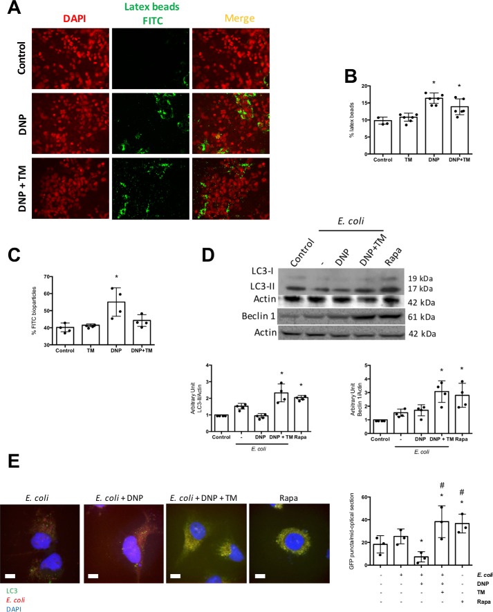
Figure 2.
Tunicamycin promotes killing of internalized bacteria and not suppression of uptake. T84 epithelial cells were incubated with fluorescent latex beads (108 1-μm diameter), DNP (0.1 mm), or DNP + TM (10 μg/ml) for 16 h, and monolayers were assessed by fluorescence microscopy (representative images, ×40 magnification) (A) and (B) flow cytometry (B). C, in parallel cultures, flow cytometry was used to assess intracellular levels of fluorescent dead E. coli F12 bioparticles (106/ml). D, T84 cells were treated with E. coli (HB101, 106 cfu) ± DNP or DNP + TM for 16 h, and autophagy was assessed by expression of LC3-II and beclin-1 on immunoblots that were quantified by densitometry and normalized to actin expression. Rapamycin (Rapa; 100 nm, 16 h) was a positive control for the induction of autophagy. E, LC3-GFP HeLa cells were treated as shown for 16 h and GFP+ puncta counted in randomly selected cells based on DAPI nuclear staining (representative images; scale bar, 65 μm). Co-localization of bacteria in phagosomes was obvious in the E. coli + DNP + TM-treated cells (mean ± S.D.; * and #, p < 0.05 compared with control and E. coli+ DNP-treated cells, respectively).

