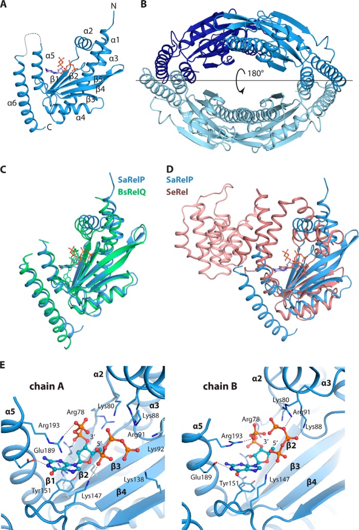
Figure 1.
Overall structure of SaRelP in the post-catalytic state. A, crystal structure of the pppGpp-bound SaRelP monomer shown in schematic with N and C termini and secondary structure elements indicated and pppGpp in ball and stick. The disordered loop 194–201 is shown with a dashed line. B, overview of the SaRelP homotetramer with the four chains in various shades of blue. The horizontal line indicates the location of the 2-fold crystallographic axis that generates the tetramer. C, structural alignment of SaRelP (blue) and BsRelQ (PDB code 5DED, green) (16). The pppGpp molecule bound at the active site in both structures is shown in ball and stick in matching colors. D, structural alignment of SaRelP (blue) with the catalytic domain of SeRel (PDB code 1VJ7, brown) (11). The GDP molecule in the SeRel active site is shown in ball and stick. The hydrolase domain of SeRel is highlighted with a darker shade of brown. E, details of the interactions between pppGpp and SaRelP in chains A and B of the structure. The pppGpp molecule is shown in ball and stick with relevant interacting residues and secondary structure elements labeled.

