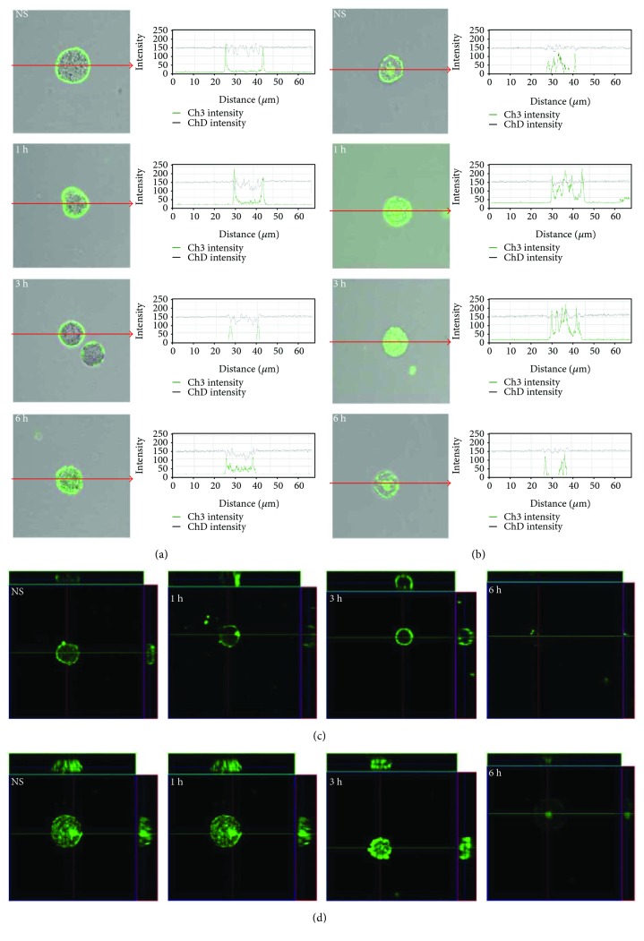
Figure 3.
Effect of LL-37 stimulation on TLR3 expression in mast cells. Mast cells were incubated with LL-37 at a final concentration of 1 μg/mL or medium alone (NS) for 1 h, 3 h, or 6 h. Representative images showing TLR3 cellular localization analyzed by confocal microscopy in (a, c) non- and (b, d) permeabilized cells. Single confocal sections (midsection of cells) reveal the surface and intracellular presence of TLR3. The signal was visualized with green Alexa 488. Fluorescence intensity diagrams showing the distribution of fluorescence in cells were mounted.

