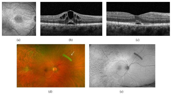
Figure 1.
(a) Infrared fundus photo of the macula showing cystoid macular edema (CME); (b) optical coherence tomography (OCT) of the macula showing CME prior to treatment; (c) macular OCT showing resolution of CME with small amount of subretinal fluid; (d) color fundus photo after resolution of retinitis with white arrow pointing to the dexamethasone intravitreal implant; (e) autofluorescence fundus photo demonstrating temporal hypofluorescent changes that represent scarring following syphilitic retinitis.

