Fig. 3.

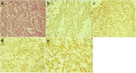
Histological staining of sections of resected tumor showing significant nuclear atypia, hyperchromasia, and pleomorphism of the tumor cells (a) and positive staining for CK-pan (b), vimentin (c), melan-A (d), and synaptophysin (e)

Histological staining of sections of resected tumor showing significant nuclear atypia, hyperchromasia, and pleomorphism of the tumor cells (a) and positive staining for CK-pan (b), vimentin (c), melan-A (d), and synaptophysin (e)