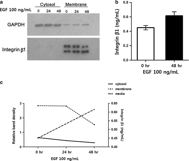
Fig. 4.

Analysis of integrin β1 localization and shedding. a Integrin β1 localization in the membrane and cytosol was examined by subcellular fractionation and western blotting. b Integrin β1 shedding was monitored by ELISA after stimulation with EGF for 48 h. c EGF-dependent changes in integrin β1 subcellular localization were examined by densitometric quantification of data shown in a
