Fig. 1.

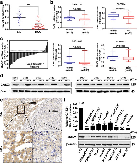
CASZ1 is downregulated in human HCC tissues and cell lines a. CASZ1 mRNA expression in 15 normal liver tissues (NLs) and 50 HCC tissues was analyzed by qRT-PCR. Data are shown as mean ± SD. ***P < 0.001. b CASZ1 expression was lower in HCC tissues than NLs according to the analysis of data from GEO (GSE62232, GSE6764, GSE25097, GSE64041, all P < 0.05). c Waterfall plot showing the downregulation of CASZ1 in 34 of 50 (68%) HCC samples compared to their matched adjacent non-tumor liver tissues (ANLTs). d The CASZ1 protein levels in HCCs, ANLTs and NLs were analyzed by Western blot. β-actin was used as a loading control. e Representative immunohistochemical images demonstrated CASZ1 protein was lowly expressed in HCC tumor tissues compared with their peri-tumor tissues. Magnification, × 100, × 400. f Expression of CASZ1 in HCC cell lines and L02, the normal liver cell line, were measured by qRT-PCR and western blot, respectively. *P < 0.05; **P < 0.01; ***P < 0.001 compared with L02
