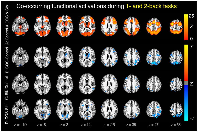
Figure 1.
Decreased functional activation in patients and siblings compared to controls during 1- and 2-back tasks. Note: Colored Z-scores indicate co-occurring significant brain activations during both 1- and 2-back tasks versus 0-back task. A typical working memory (WM)-related activation pattern was found in patients with childhood-onset schizophrenia (COS), their siblings, and healthy controls as shown in the union activation map of these three groups (A). Within these activated regions, both patients (B) and siblings (C) showed reduced activations compared with controls. In addition, patients showed lower activations than their siblings (D). All clusters are significant (p < .05, corrected), and co-occurring maps were generated by minimum Z conjunction of 1- and 2-back contrasts. Montreal Neurological Institute coordinates of peak regions in (B), (C), and (D) are listed in Table S2, available online.

