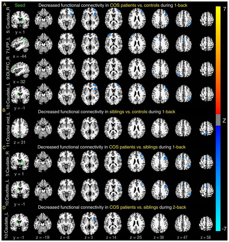
Figure 2.
Decreased functional connectivity in patients compared to controls and siblings. Note: Patients showed significantly lower functional connectivity between seeds in the left column and many of brain regions on the right (that were activated, shown in Figure 1A) than controls during 1-back (A) and siblings during 1-back (C) and 2-back (D). In addition, siblings also showed lower functional connectivity than controls (B). Seeds were defined by regions showing lower activations in patients or siblings compared with controls (see details in Figure S6, available online). All clusters are significant (p < .05, corrected for both the number of voxels and seeds examined). LPP = lateral posterior parietal cortex.

