Figure 6.
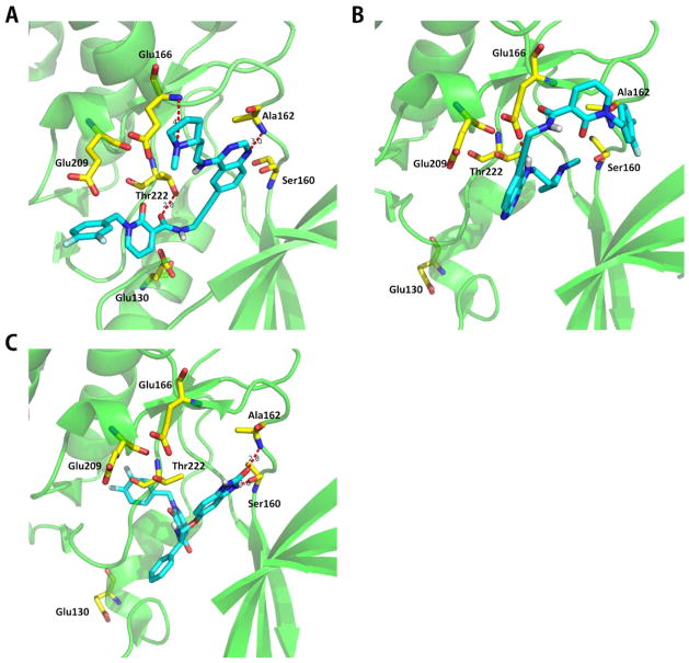
Compound CHEMBL3640476 bound to the PDPK1 ATP binding site. Structures of the compound CHEMBL3640476 binds to the PDPK1 ATP binding site in the kinase domain. (A) CHEMBL3640476 binds to the PDPK1 crystal structure 3NAY (docking score 8.49). (B) CHEMBL3640476 binds to the PDPK1 crystal structure 3NAX (docking score 5.35). (C) Original structure of 3NAX (PDPK1 cocrystallized with compound 7). Blue sticks represent the ATP competitive inhibitor of PDPK1, named CHEMBL3640476. Yellow sticks represent the important residues of PDPK1 ATP binding domain which are also labeled. H-bonds to the important residues are displayed as dashed red lines. In 3NAY, CHEMBL3640476 has H-bonds to Ala162, Glu166, and Thr222 within the ATP-site of PDPK1. Glu130 is also in the active binding site. However, only one H-bond to Glu166 is formed while Glu130 is out of the active site in the 3NAX.

