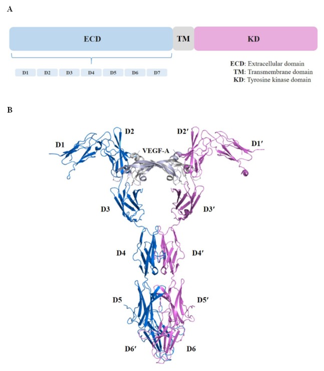
Fig. 1.
Structure of the VEGFR-1 extracellular domain in complex with VEGF-A. (A) Schematic representation of the domain organization of VEGFR is shown. (B) Complex crystal structure of VEGFR-1 extracellular domain with VEGF-A (PDB ID: 5T89) is shown. We have shown the structure in a ribbon representation with each chain depicted by a different color. The chains of the VEGF-A homodimer are shown in light blue and gray, and the VEGFR-1 D1–D6 chains in deep blue and magenta.

