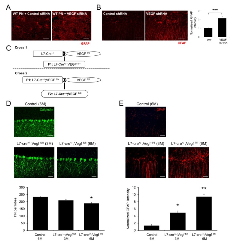
Fig. 2.
Effect of PNs specific deletion of VEGF on PNs survival and neuroinflammation. (A) Representative confocal images of astrocytes activation in cultured cerebellar cells after VEGF siRNA treatment. Scale bar, 50 μm. (B) Representative fluorescence images and quantification of GFAP in VEGF shRNA treated mice cerebellum (n = 3 per group). Scale bars, 50 μm. (C) Generation of L7/Pcp2-cre;VEGFflox/flox mice. (D) Confocal images and quantification of cerebellar PNs in control and L7/Pcp2-cre;VEGFflox/flox mice (IV and V lobes, n = 3–4 per group). Scale bars, 50 μm. (E) Representative confocal images and quantification of astrocytes activation in control and L7/Pcp2-cre;VEGFflox/flox mice (n = 3–4 per group). Scale bars, 50 μm. (B, D, E) Student’s t test. *P < 0.05, **P < 0.01, ***P < 0.001. All error bars indicate standard error of the mean.

