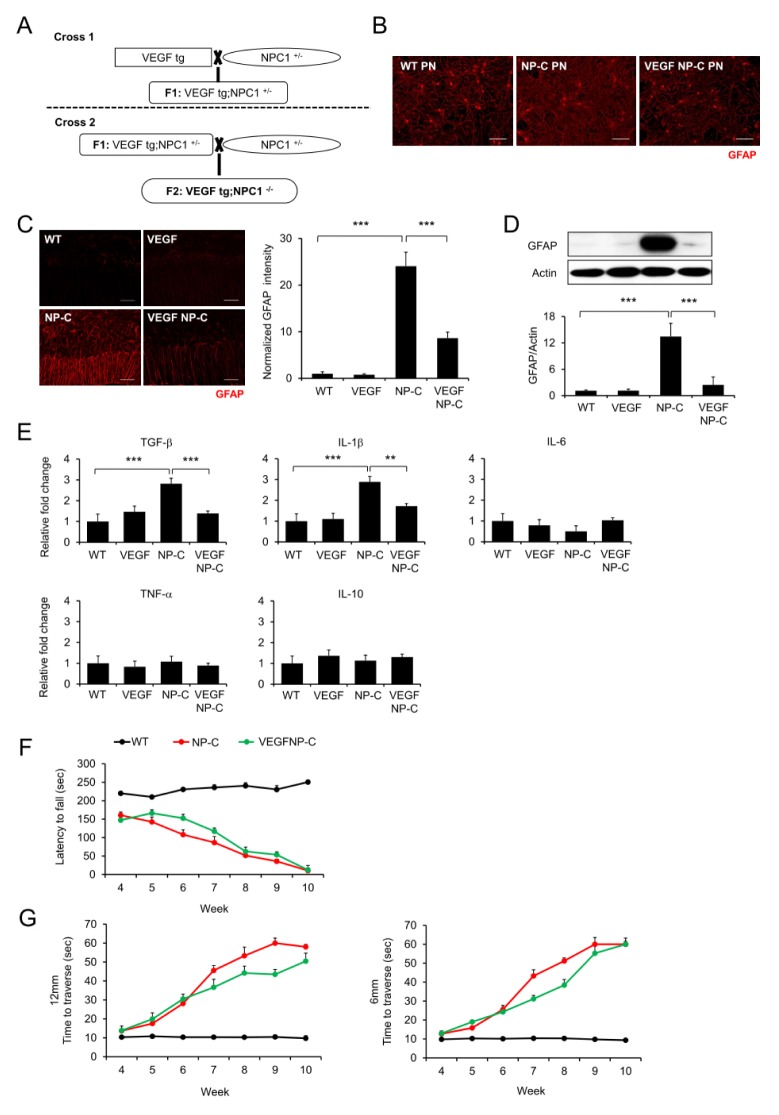
Fig. 3.
Improvement of NP-C pathology by replenishment of VEGF. (A) Generation of VEGF/NP-C mice. (B) Representative confocal images of astrocytes activation in cultured cerebellar cells derived from wild-type (WT), NP-C, VEGF, and VEGF/NP-C mice. Scale bar, 50 μm. (C) Representative fluorescence images and quantification of GFAP in cerebellum of wild-type (WT), NP-C, VEGF, and VEGF/NP-C mice (n = 3 per group). Scale bars, 50 μm. (D) Western blot analysis of GFAP in cerebellum of WT, NP-C, VEGF and VEGF/NP-C mice (n = 3 per group). (E) mRNA levels of inflammation related genes in the cerebellum of WT, NP-C, VEGF and VEGF/NP-C mice (n = 3 per group). (F) Rota-rod scores of WT, NP-C and VEGF/NP-C mice (n = 6–7 per group). (G) Beam test of WT, NP-C and VEGF/NP-C mice. Left, 12-mm square beam. Right, 6-mm square beam (n = 6–7 per group). (C-G) One-way analysis of variance, Tukey’s post hoc test. **P < 0.01, ***P < 0.001. All error bars indicate standard error of the mean.

