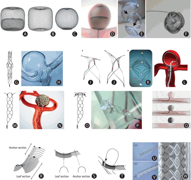
Figure 2.
New devices for intracranial aneurysm treatment. (A-D) Woven Endo-Bridge (WEB) devices: (A) a double-layer, (B) a single-layer, and (C) a single-layer, sphere-shaped WEB device; (D) a WEB double-layer device deployed from a catheter into a bifurcation aneurysm (Copyright Sequent Medical). (E, F) Medina embolization device (MED): (E) deployment of the MED showing the petals of the device (arrows), and (F) fully deployed device showing its three-dimensional conformation (Copyright Medtronic). (G, H) The Barrel stent device: (G) the original shape of Barrel stent, and (H) the bulged center section of the stent provides complete neck coverage in a flow model (Copyright Medtronic). (I-L) The PulseRider device: (I) “T” configuration, (J) “Y” configuration, (K) view from above; (L) PulseRider deployment at the neck of a bifurcation aneurysm (Copyright Pulsar Vascular). (M, N) The pCONus: (M) the pCONus has a stent-like proximal shaft with four distal petals; (N) the four distal loops of the pCONus are deployed inside the aneurysm at the neck level, assisting coil occlusion. (O, P) The pCANvas, an evolution of the pCONus with additional membrane coverage of the petals (arrows in O, Copyright Phenox); (Q) the Comaneci device. The images show the ‘deflated’ and ‘inflated’ status of Comaneci, as well as after removal of the device. (R-T) The eCLIPs: (R, S) the eCLIPs device has a leaf section and an anchor section; (T) deployment position of the eCLIPs device, with the leaf section covering the bifurcation aneurysm and allowing microcatheter access to the aneurysm sac (Copyright Evasc Medical Systems Corp.). (U-W) The balloon-expandable honeycomb microporous covered stent crimped on the balloon catheter (U), completely expanded (V), and removed from the catheter (W). Adapted from Nakayama et al., with permission from Springer Nature [45].

