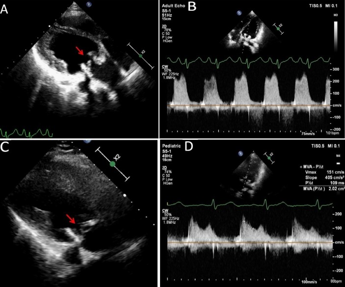
Figure 2.
Echocardiogram recorded at presentation shows increased thickness of the bioprosthetic valve leaflets (A) (arrow). The pressure gradient across the mitral bioprosthesis is increased significantly (B). Echocardiogram recorded after the treatment with corticosteroids and anticoagulation (C,D). There is remarkable thinning of the bioprosthetic valve leaflets (C) (arrow). The pressure gradient across the mitral bioprosthesis has decreased significantly (D). MVA, mitral valve area.

