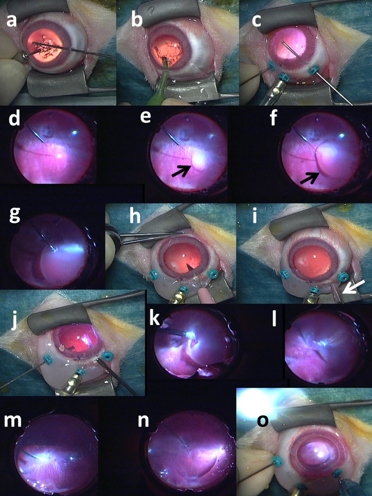
Fig. 1.
Surgical procedures to implant retinal prosthesis, OURePTM, in right eye of rhodopsin-transgenic white rabbit. a) Lens anterior capsule is cut with 25G vitreous cutter under irrigation with 25G infusion cannula in anterior chamber. b) Lens nucleus and cortex is aspirated with phaco-tip from corneal incision. c) Three 25G trocars are inserted over conjunctiva through sclera into vitreous at 2.5 mm from corneal limbus: a middle trocar is connected with infusion cannula, and the other two trocars are used for vitreous cutter and light guide. Posterior capsule is cut with vitreous cutter. d) After vitreous gel has been cut, subretinal fluid infusion is started with 38G tip. e) Bleb retinal detachment (arrow) is made by 38G tip infusion of BSS-Plus solution. f) Bleb (arrow) is enlarged with further infusion. g) Retinal tear is made by retinal coagulation with 25G bipolar diathermy. h) Scleral incision is made with 22.5° knife after conjunctival incision. i) Rolled-up dye-coupled film (arrow) is inserted through scleral incision with 20G subretinal forceps. j) Rolled-up film is inserted into vitreous with 20G subretinal forceps. k) Rolled-up film is inserted into subretinal space through retinal tear with 20G subretinal forceps. l) Film is now under detachment retina. m) Fluid-air exchange in vitreous cavity is accomplished with 25G vitreous cutter in aspiration mode to reattach the retina. n) Laser photocoagulation is applied around retinal tear. o) Silicone oil is injected in vitreous cavity with 25G tip. Finally, scleral and conjunctival incision is sutured and trocars are removed (not shown).

