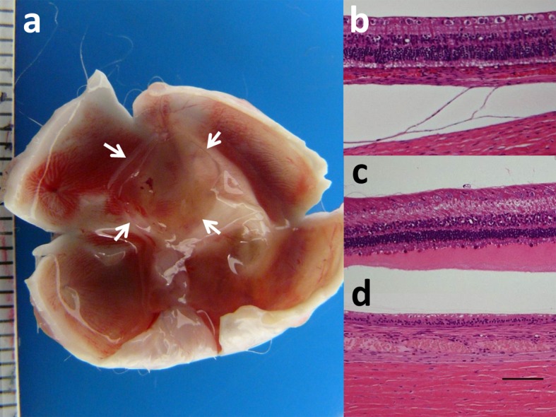
Fig. 2.

Macroscopic view of unfixed and dissected eye with subretinal square dye-coupled film (5 × 5 mm, arrows) in 1-month implantation (a). Light microscopic sections of the retina of the posterior pole near film implantation in normal rabbit with 1-month film implantation (b), rhodopsin-transgenic rabbit with 1-month film implantation (c), and normal rabbit with 1 month observation after removal of 6-month implanted film (d). The photoreceptors are at bottom of photographs. Separation between the choroid and sclera is artifact (b). Photoreceptor outer segments are relatively maintained in normal rabbit (b) while outer segments are shortened with eosin-stained serous fluid in rhodopsin-transgenic rabbit (c). Loss of retinal outer nuclear layer is noted 1 month after removal of 6-month implanted film in normal rabbit (d). Hematoxylin-eosin stain. Scale in ruler is 1 mm in a. Scale bar=100 µm in b, c and d.
