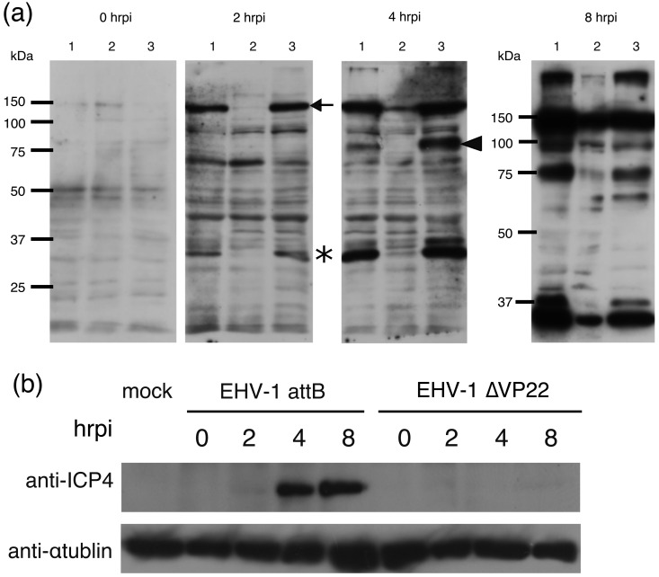
Fig. 2.
Protein expression comparison by Western blotting using anti-EHV-1 antibody (a). MDBK cells were infected with EHV-1 attB (lane 1), EHV-1∆VP22 (lane 2) and EHV-1∆VP22R (lane 3) at an MOI of 3 PFU/cell and analyzed by Western blotting with antibody to EHV-1 virions at 0, 2, 4 and 8 hrpi. Molecular sizes are shown on the left (a). Expression of ICP4s of EHV-1 attB, EHV-1∆VP22 and EHV-1∆VP22R were analyzed by Western blotting using anti-ICP4 antibody (b).

