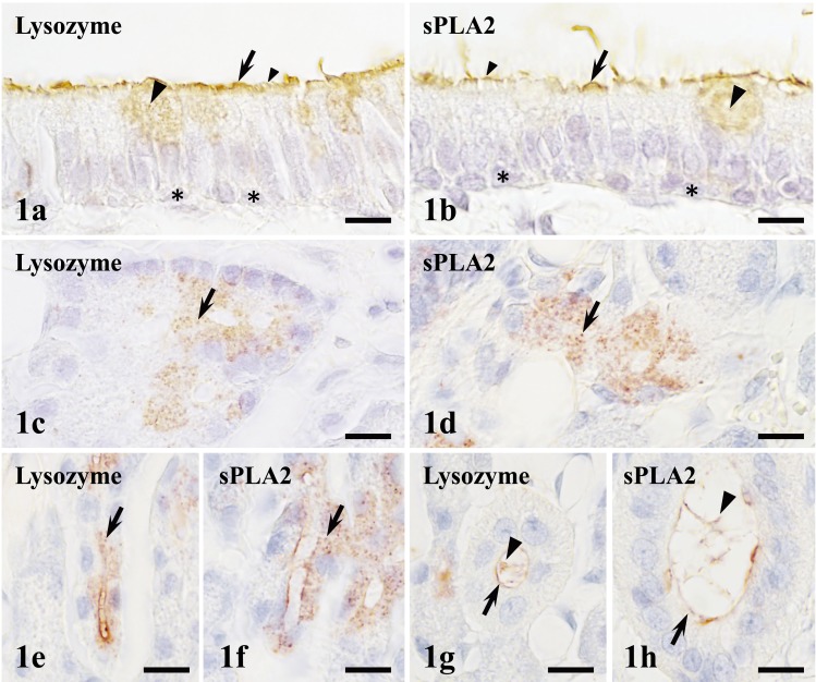
Fig. 1.
The localization of lysozyme and sPLA2 in the nasal mucosa. In nasal epithelium, lysozyme and sPLA2 are immunopositive in the cytoplasm of NC (arrows in a and b), secretory granules of GC (large arrowheads in a and b) and cilia of ciliated epithelial cells (small arrowheads in a and b), but negative in cytoplasm of ciliated epithelial cells and basal epithelial cells (asterisks in a and b). In the nasal gland, lysozyme and sPLA2 are immunopositive in secretory granules of serous acinar cells (arrows in c and d) and intercalated ducts (arrows in e and f). Immunopositivities of lysozyme and sPLA2 are also visible on the luminal surface of cells of exocrine ducts (arrows in g and h) and luminal contents (large arrowheads in g and h). Bar=10 µm.

