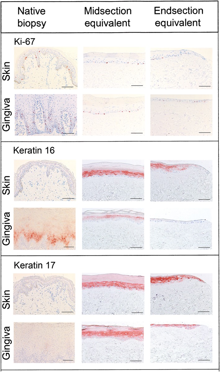
Figure 3.

Histology and immunohistochemistry of native skin and gingiva biopsies and skin and gingiva substitutes. Representative Ki‐67 and keratin 16 and 17 staining is shown for native biopsy tissue, the midsection and the migrating front (end section) of skin and gingiva substitutes. Scale bar = 100 μm
