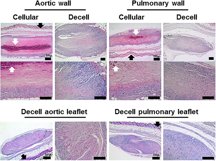
Figure 5.

Images of sections of explanted cellular and decellularized aortic and pulmonary valve tissues (stained with H&E) 12 weeks after subcutaneous implantation in mice. Samples of cellular and decellularized aortic and pulmonary artery tissues from the same valved conduits were implanted subcutaneously in mice for 12 weeks. Both the cellular aortic and pulmonary artery show evidence of capsule formation, limited cell infiltration and areas of calcification and tissue deterioration. The explanted decellularized aortic and pulmonary arterial wall tissues show high numbers of cells infiltrating the peripheral regions of the implants with cells penetrating the central regions and little evidence of tissue calcification or deterioration. Samples of decellularized pulmonary and aortic valve leaflet tissues implanted subcutaneously in mice for 12 weeks were fully infiltrated with cells with no evidence of tissue deterioration or calcification. Black arrows indicate mouse epidermis. White arrows indicate areas of calcification. Occasional black spots are sectioned mouse hair. Scale bars 200 μm unless otherwise labelled [Colour figure can be viewed at wileyonlinelibrary.com]
