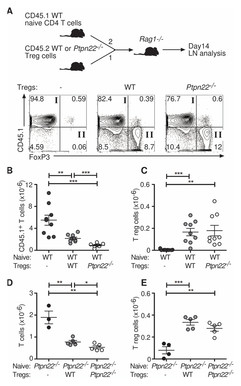
Fig. 3. Ptpn22-/- Tregs are more suppressive than WT Tregs in vivo.
(A to C) WT naïve CD45.1+CD4+CD25-CD44lo cells (4 x 10⁵) were injected alone or together with WT or Ptpn22-/- CD45.2+CD4+CD25+ Tregs (2 x 10⁵) i.v. into Rag1-/- recipients in a 2:1 ratio as indicated (n = 9 mice per group). After 14 days, lymph nodes were assessed by flow cytometry for their proportions of CD45.1+FoxP3- cells (quadrant I) and CD45.1-FoxP3+ Tregs (quadrant II). Representative plots are shown from each group. Absolute numbers of (B) CD45.1+FoxP3- cells and (C) CD45.1-FoxP3+ Treg are shown. Each symbol represents an individual mouse. Bars represent SEM. Data are representative of 3 individual experiments. (D and E) Ptpn22-/- naïve CD4+CD25-CD44lo cells (1 x 10⁵) were injected alone or together with WT or Ptpn22-/- thymic CD4+CD25+ Tregs (0.25 x 10⁵) i.v. into Rag1-/- recipients in a 4:1 ratio (n = 3-5 mice per group). After 19 days, lymph nodes were assessed for the numbers of (D) FoxP3- cells and (E) FoxP3+ Tregs. Each symbol represents an individual mouse. Bars represent SEM. Data are representative of 2 individual experiments. **P < 0.005, ***P < 0.0005.

