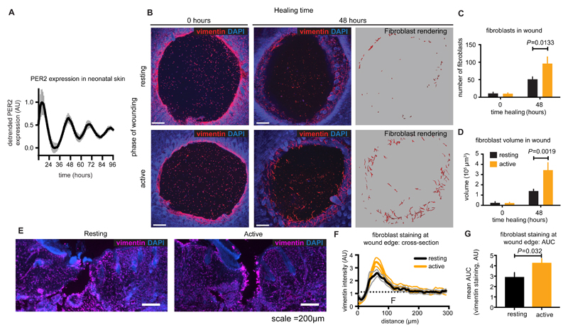
Fig. 5. Diurnal variation in wound healing outcome and fibroblast mobilisation.
A. Bioluminescent recording of PER2 expression in neonatal (P5) skin explants from PER2::LUC mice (mean±SEM n=6). B. Mouse skin wounds before and after 48 hours of healing. Fibroblasts were identified by anti-vimentin reactivity (red) and morphology, and quantified by number (C ) and volume (D) (mean±SEM, n=6-7, Holm-Sidak’s adjusted P value is indicated). Scale bar =200 μm. E. 60 μm transverse sections of mouse wounds made during the active and resting phases stained using anti-vimentin (magenta) and Hoescht (blue). Cross-sectional vimentin staining across wound edges was quantified (F, mean±SEM) and Area Under Curve (AUC) was calculated using distal vimentin as a baseline (G) (mean±SEM, n=16 (active) or 20 (resting), P from a student’s t test is indicated).

