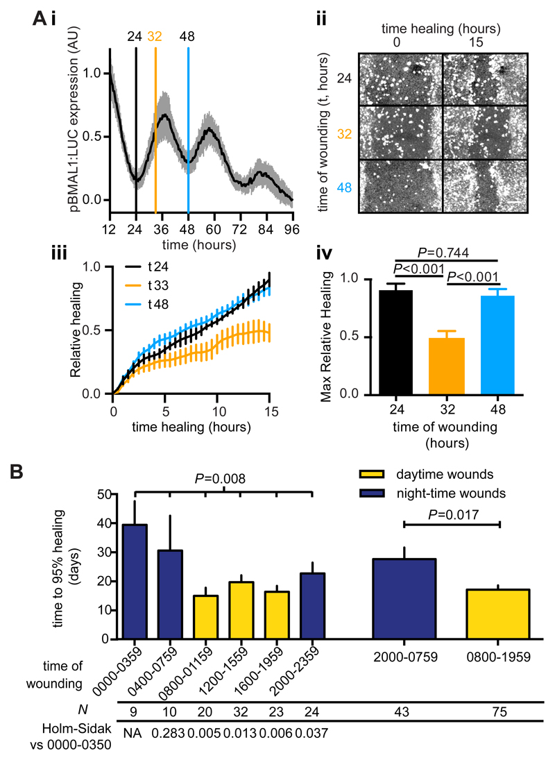
Fig. 6. A circadian rhythm in keratinocyte wound healing and a diurnal variation in human burn healing outcome.
A. Synchronised human HaCaT keratinocyte monolayers expressing luciferase under control of the BMAL1 promoter (i, mean±SD, n=24) were wounded at the indicated times (vertical lines) and healing monitored by confocal microscopy (ii). Relative fluorescence in the wound area (iii, mean±SEM n=4) was calculated and maximal healing after 15 hrs was compared by Tukey’s multiple comparisons test (iv, P values are indicated) B. Mean time to 95% healing ±SEM from 118 human burn incidents separated by time of burn occurrence in 4 (left) or 12 hour (right) bins. ANOVA P value is indicated, as is the P value for Welch’s t-test comparing daytime vs night-time wounds. P values from Holm-Sidak’s test versus the 0000-0359 bin are indicated below.

