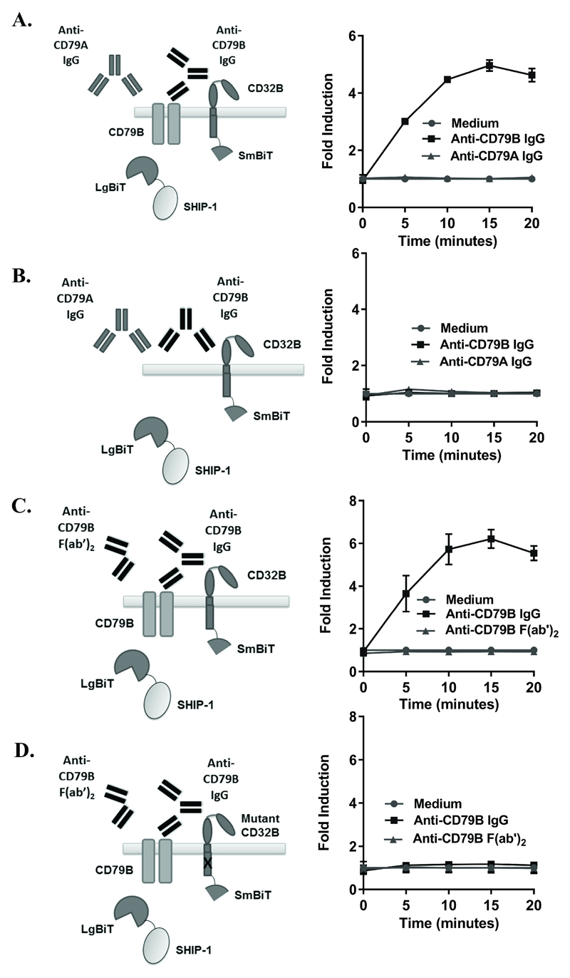
Figure 3. Detection of SHIP-1 recruitment following CD32B-CD79B crosslinking.
(A,B) HEK293F cells, transiently co-transfected with CD32B-SmBiT, SHIP-1-LgBiT and CD79B (A) or CD32B-SmBiT, SHIP-1-LgBiT alone (B) on d0 were stimulated with anti-CD79B IgG (AT105-1) or anti-CD79A IgG (ZL7-4) (20 µg/ml) on d1. Luminescence was measured at time 0 following substrate addition, and at 5 minute intervals following stimulation. (C, D) HEK293F cells, transiently co-transfected as in A) (C), or with ITIM-mutated CD32B-SmBiT, SHIP-1-LgBiT and CD79B (D) on d0 were stimulated with anti-CD79B IgG (ZL9-3 IgG) or F(ab’)2 (ZL9-3 F(ab’)2) (10 µg/ml) on d1. Luminescence was measured as above. (A-D) Left – schematic diagrams; right – fold inductions. Means ± S.D. of technical replicates are shown, representative of 5 (A) and 4 (C) independent experiments. (B, D) Control experiments performed at the same time as A) and C) respectively, representative of 3 independent experiments.

