
Figure 5.
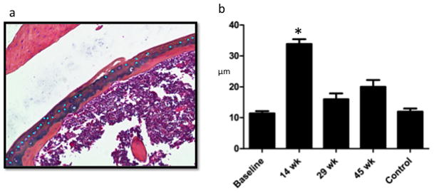
Number of Mitotic Figures is Significantly Greater During Proliferative Period (14 weeks). a. Sample mitotic figure measurement during proliferative period: using a touchscreen pen, mitotic figures were marked superficial to the mineralized cartilage (within the articular cartilage) within one standardized 10× power field (bright blue dots). Each investigator selected their own field with the goal to include maximal articular cartilage area. b. Osteomeasure counted the number of marks made by each investigator and means were calculated showing significantly more mitotic figures during the proliferative period.