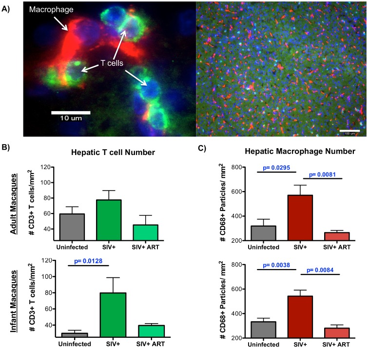
Fig 1. Increased number of macrophages and T cells observed in the liver during SIV infection.
CD68 macrophages and CD3 T cells were enumerated in the liver of uninfected, chronically SIV-infected, and chronically SIV-infected, cART infant or adult rhesus macaques by immunofluorescence microscopy. A) Fluorescent images at 600x (left image, scale bar = 10 um) and 100x (right image, scale bar = 100 um) depicting specific staining for macrophages (red) and T cells (green) in the liver (blue indicates nuclei). For quantification, eight random fields in the liver were imaged under 100x magnification and then analyzed by ImageJ software. B) Quantification of eight random fields of view for each animal to enumerate CD3 T cells by ImageJ Cell Counter analysis with adult and infant macaques graphed separately (top and bottom panels, respectively). C) Quantification CD68 macrophages in eight random fields of view for each animal using ImageJ Particle analysis. Data are graphed as the mean ± SEM. Statistical significance between groups was determined Mann Whitney T tests.

