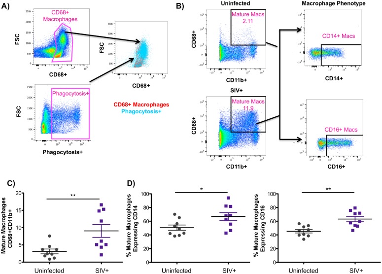
Fig 4. Evaluation of CD68+ mature macrophages in the liver.
A) Freshly isolated macaque liver cells were incubated with fluorescently labeled E. coli bioparticles for 2 hours. After washing, cells were evaluated for phagocytosis of E.coli by flow cytometry. Gating first on single, live, CD45+, CD3- cells, CD68+ liver cells were found to be the primary cell subset to phagocytosis E. coli. B) Liver cells suspensions were first gated on single, live, CD45+, CD3- cells. Mature macrophages, identified as CD68+CD11b+, were evaluated for the expression of CD14 and CD16. C) Frequency of mature CD68+CD11b+ macrophages in the liver of uninfected (gray symbols) and SIV-infected (purple symbols) macaques. D) Phenotype of mature hepatic macrophages in uninfected (gray symbols) and SIV-infected (purple symbols) macaques based on CD14 and CD16 expression.

