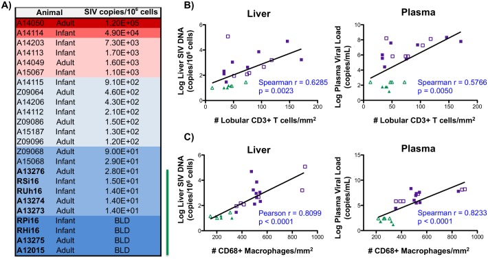
Fig 6. Liver macrophage number correlates with SIV levels.
A) SIV DNA copies in samples from the livers of both infant and adult macaques were determined by quantitative hybrid real-time/digital PCR and normalized per million cell equivalents, based on parallel analysis of a single copy rhesus macaque CCR5 target template. Macaques treated with cART are denoted with bolded font and a green line to the right of the table (BLD = below the limit of detection). B-C) Correlation analysis between copies of SIV DNA in the liver (left) or plasma SIV levels (right) and liver T cell number (B) or liver macrophage number (C) in SIV-infected (purple symbols) and SIV-infected cART (green symbols) macaques. For macaques having SIV plasma viral load and SIV DNA copies below the limit of detection, the log transformed limit of detection values (30 copies/mL for plasma or 10 copies/106 cells for liver) were used for correlation analysis. Adult (open symbols) and infant (closed symbols) macaques are denoted.

