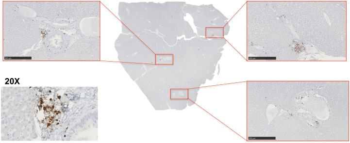
Fig 7. SIV-infected cells localize around the portal triads.
A) RNAscope in situ hybridization was used to detect SIV RNA-positive cells in the liver. Images were captured from whole tissue scans at 5x magnification (images outlined in red, scale bar = 500 um) and depict SIV-infected cells around the portal triads. A representative 20x image of a portal triad was included of the SIV-RNA positive cells (brown cells).

