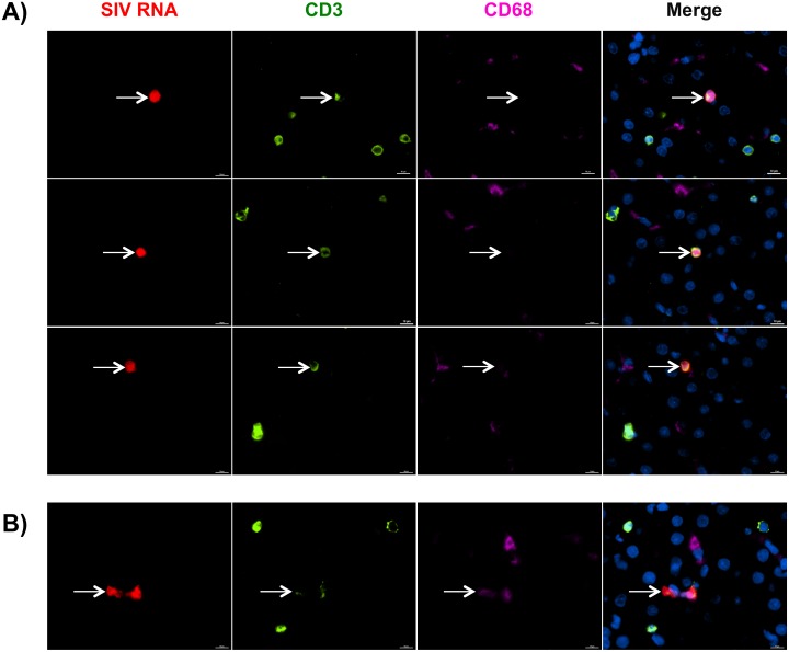
Fig 8. T cells are the primary cellular subset infected with SIV in the liver.
A-B) Liver tissue sections from SIV-infected untreated macaques were assessed for SIV RNA+ cells (red) by in situ RNAscope technology followed by antibody staining with CD3 to identify T cells (green) and CD68 to identify macrophages (pink). Nuclei were stained with Dapi (blue). SIV RNA+ signal was predominately found associated with CD3 T cells (A) and in rare cases CD68 macrophages (B).

