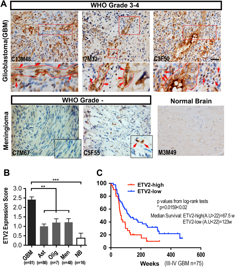
Fig. 2. ETV2 expression is preferentially detected in high-grade human gliomas.
a IHC staining for ETV2 of human brain tumors and normal brain tissues (GBM grade III–IV, n = 81; astrocytoma grade I–II, n = 86; meningioma grade I–II, n = 48; oligodendroglioma grade I–II, n = 7 and normal brain tissues, n = 5); areas in dashed boxes are magnified below; ETV2+ cells were highlighted by dashed circles or arrows. See also Fig. S1. Scale bar: 50 μm. b Quantitative analysis of the expression level of ETV2 in different human brain tumors and normal tissues. c Kaplan–Meier analysis of patient OS according to the ETV2 expression level in high-grade brain tumors (n = 77). From the TCGA database, a total of 77 high-grade brain tumor patients were divided into ETV2-high (n = 30) or ETV2-low (n = 47) groups, in which the cutoff value was the average ETV2 expression level of all patients

