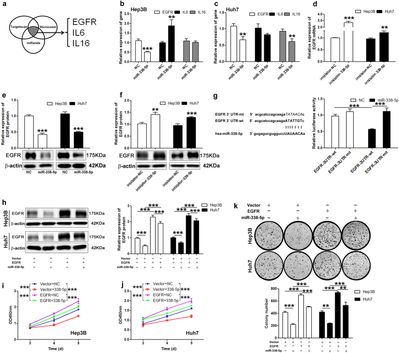
Fig. 4. EGFR is another direct target of miR-338-5p.
a The target genes of miR-338-5p were predicted using TargetScan, MicroCosm, and MiRanda. b–c The mRNA levels of potential target genes of miR-338-5p were measured after cells were transfected with miR-338-5p for 72 h. d EGFR mRNA expression was detected after cells were treated with miR-338-5p inhibitor by qRT–PCR. The protein expression of EGFR in HCC cells transfected with miR-338-5p mimics e or inhibitors f was analyzed by western blotting. g Left: sequence complementarity between the 3′-UTR of EGFR mRNA and the seed region of miR-338-5p. Right: luciferase activity in Hep3B cells transfected with miR-338-5p and reporter plasmids containing wt (wild-type) or mt (mutant) EGFR 3′-UTR was analyzed. h Cells were treated with the EGFR vector (EGFR, hereafter), and then, the protein levels of EGFR were measured by western blotting. The proliferation i–j and colony formation k of Hep3B and Huh7 cells were remarkably promoted after transfection with the EGFR vector compared with the control vector. Student’s t-test (two-tailed) or one-way or two-way analysis of variance was employed to analyze the data. Data in all experiments are presented as the means ± SD of three independent experiments. **P < 0.01, ***P < 0.001 vs. NC

