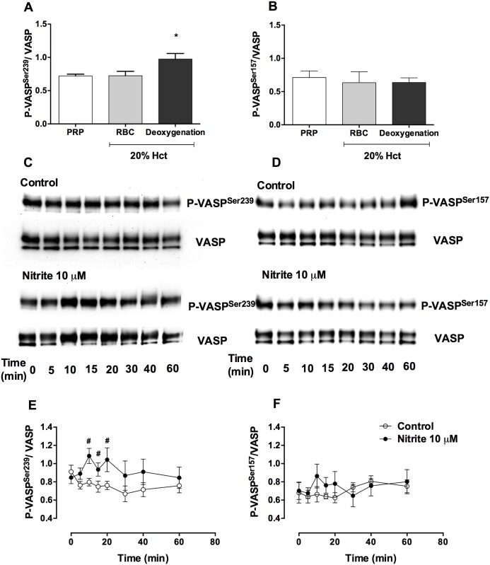
Fig 2. Nitrite increased P-VASPSer239 but not P-VASPSer157 in the presence of deoxygenated erythrocytes.
Quantification of baseline P-VASPSer239/ VASP (A) and P-VASPSer157/ VASP (B) in platelets from PRP, PRP + erythrocytes (20% hematocrit) with or without deoxygenation, representative Western blot bands of P-VASPSer239 (C), P-VASPSer157 (D), VASP expression and quantification of P-VASPSer239/VASP (E) and P-VASPSer157/VASP (F) in control and nitrite treated deoxygenated PRP + erythrocytes (20% hematocrit) at different time points are shown in this figure. PRP + erythrocytes (20% hematocrit) were deoxygenated by helium for 10 minutes (pO2 ~ 25 mmHg). Nitrite (10 μM) was added in deoxygenated samples and incubated for 5, 10, 15, 20, 30, 40 and 60 minutes. Data are mean ± SEM (n = 7). *P < 0.05 compared with PRP and tested by one-way ANOVA with Tukey’s multiple comparison. #P < 0.05 compared with control and tested by paired Student’s t-test.

