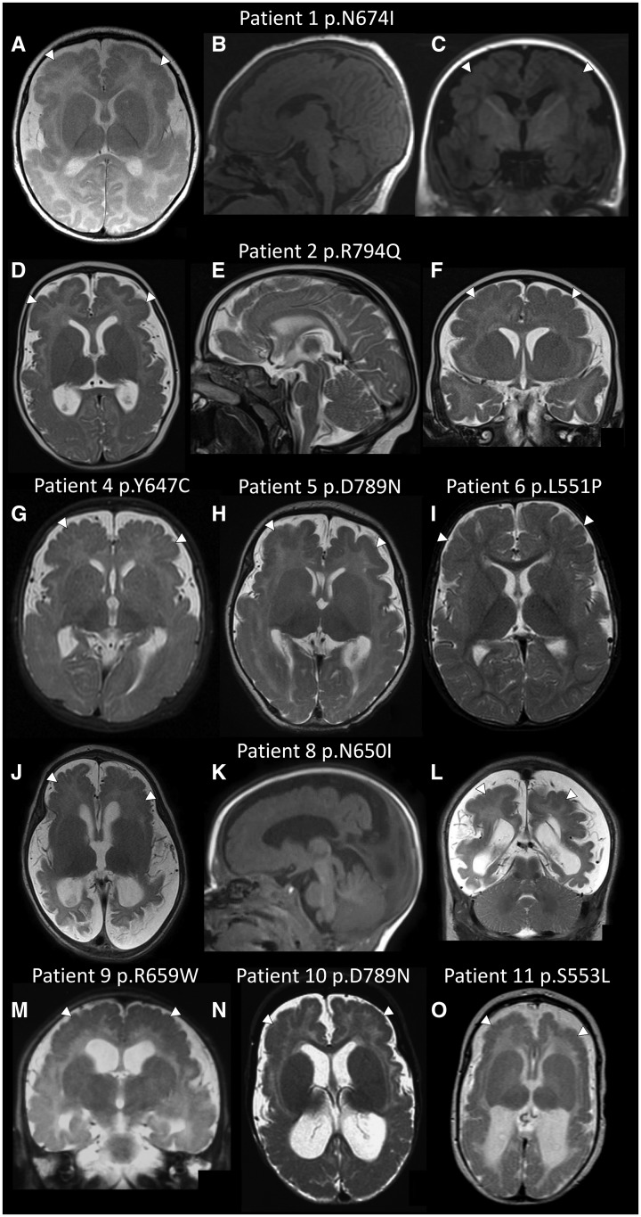
Figure 1.
Polymicrogyria in patients with GRIN1 mutations. Axial, midline sagittal and coronal brain magnetic resonance images for Patient 1 at age 2 months (A–C) and Patient 2 at age 5 months (D–F); axial magnetic resonance images for Patient 4 at age 3 months (G), Patient 5 at age 6 weeks (H) and Patient 6 at age 8 months (I); axial, sagittal and coronal images for Patient 8 at age 3 months (J–L); a coronal image for Patient 9 at age 4 months (M); axial images from Patient 10 at age 8 months (N) and Patient 11 at age 2 months (O). Images B, C and K are T1-weighted. All other images are T2-weighted. The images demonstrate bilateral extensive polymicrogyria (white arrows) more severe anteriorly. Note the increased extra-axial spaces and enlarged lateral ventricles (in most images apart from I) suggesting cerebral volume loss.

