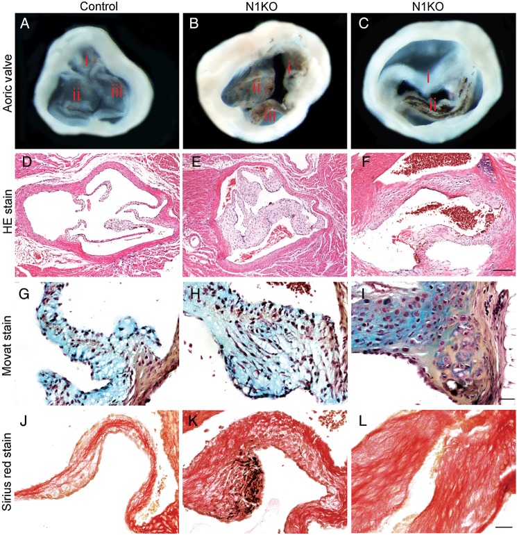
Figure 2.
Inactivation of Notch1 in valvar endothelial cells results in congenital anomalies of aortic valves and valvar aortic stenosis. Gross view of aortic valves from 4-month-old mice shows that, compared with the controls (A), N1KO mice have thickened aortic valves with pigmented nodules at the edge of leaflets (B, n = 16), and ∼30% of N1KO mice have bicuspid aortic valves (C, n = 5). Haematoxylin-and-eosin-stained (HE) sections of aortic valves shows that N1KO mice (E and F) have thickened leaflets compared with the controls (D). Movat's staining shows increased presence of glycosaminoglycan (blue) and collagen (yellow) at the base of BAV of N1KO mice (I) compared with tricuspid aortic valves (TAV) of control (G) or N1KO mice (H). Sirius red staining shows increased staining for fibrotic cells within aortic valves of N1KO mice (K and L) compared with the controls (J). Bar = 100 µm (D–F) or 20 µm (G–L).

