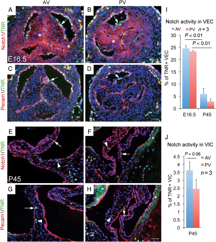
Figure 4.
Notch activity in embryonic and adult arterial valves revealed by transgenic Notch reporter mice. (A and B) Simultaneous view of the canonical Notch activity (green) and membrane Notch1 protein expression (red) in E16.5 arterial valves shows membrane Notch1 proteins in valvar endothelial cells (arrow) and valvar interstitial cells (asterisk), but Notch activity is only present on the surface of valves (arrow). (C and D) Co-staining of Pecam1 (red) and transgenic Notch reporter (green) confirms valvar endothelial cells-specific Notch activation in embryonic valves (arrow). (E–H) Immunostaining shows that membrane Notch1 proteins are predominantly present in valvar endothelial cells (arrow) of arterial valves at postnatal (P) 45 day, and active Notch signalling is seen in a small set of valvar endothelial cells and valvar interstitial cells (arrowhead). Asterisks in E and H indicate background of auto-fluorescence associated with elastic fibres. (I) and (J) show quantitative values. Bar = 50 µm.

