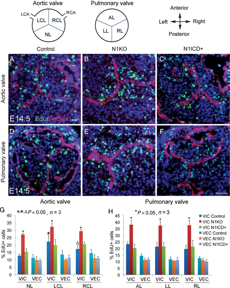
Figure 5.
Notch1 in valvar endothelial cells negatively regulates proliferation of valvar interstitial cells during development of arterial valves. Schematic diagram shows transverse view of arterial valves. LCA/RCA, left/right coronary artery; NL, non-adjacent leaflet; LCL/RCL, left/right coronary leaflet; AL/LL/RL, anterior/left/right leaflet. EdU labelling shows proliferating cells (green) in arterial valves of E14.5 control (A and D), N1KO (B and E), and N1ICD+ embryos (C and F). Pecam1 staining marks valvar endothelial cells (red). Bar = 50 µm. (G) and (H) show quantitative results. The data are presented as the ratio of EdU+ cells/total valvar endothelial cells or valvar interstitial cells. Statistical comparison was performed using one-way analysis of variance followed by Tukey's test. A P-value of <0.05 was considered significant. Asterisk indicates comparison with the control in each individual leaflet, hash indicates comparison with the non-adjacent leaflet in each genotype, and open triangle indicates comparison with the left coronary leaflet in each genotype.

