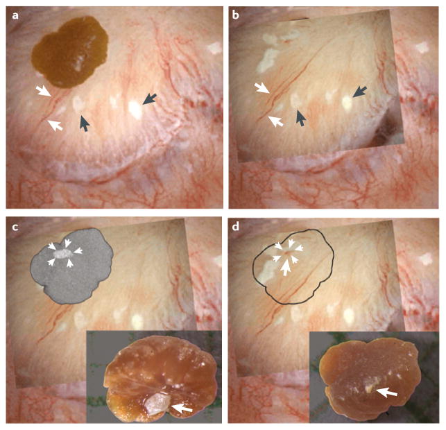
Figure 1. Interstitial plaque and corresponding attachment site on a CaOx stone.
a | Digital endoscopic image of a 3 mm CaOx stone attached to a papillum before stone removal by percutaneous nephrolithotomy. Several sites of interstitial plaque (arrowheads) are visible as well as blood vessels (arrows) that were used for image orientation. b | Following stone removal the papillum was reimaged — an overlay showing the sites of interstitial plaque and blood vessels after stone removal has been placed over the original endoscopic imagec | “Ghosted” CT image of the detached stone showing a site of calcium phosphate (arrowheads) on the papillary surface of the stone. The insert shows a light microscopic image of the papillary surface of the detached stone with the site of calcium phosphate (arrow). d | The site of calcium phosphate on the papillary surface of the stone (arrows) aligns with a region of interstitial plaque with a central blood spot (arrow) on the papillium, which is presumably the site of stone attachment. The insert shows the urinary surface of the detached stone.

