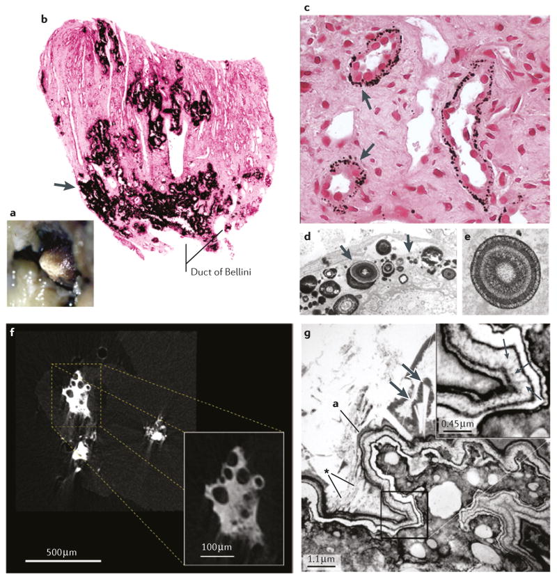
Figure 2. Sites of interstitial plaque in idiopathic CaOx stone formers.
a | Endoscopic image showing irregular whitish regions of interstitial plaque covering the papillary tip. b | A biopsy from this region showing black Yasue-stained material (arrow) within the interstitial space of the inner medulla. c | Light microscopy and d | transmission electron microscopy images showing regions of plaque in the basement membrane of the thin loops of Henle only, suggesting that plaque originates at this site. e | The plaque is composed of laminated spheres with up to nine alternating light (hydroxyapatite) and dark (matrix) layers. f | Micro CT image of plaque that has filled the interstitium and generated islands that encompass nearby tubules with no evidence of intraluminal deposits. g | Transmission electron microscopy imageshowing a multilayered ribbon-like structure separating a region of interstitial plaque (lower right) from an overgrowth of developing stone (upper left). In the thickest region of the white lamina of the ribbon (insert), tiny thin spicules run perpendicular to the surface adjacent to voids containing tightly packed crystals (small arrows). Numerous small crystals have grown into the outer border of the ribbon (asterisk) and merged with large crystals within the urinary space at the developing overgrowth. The double arrows indicate a large in-growing crystal.

