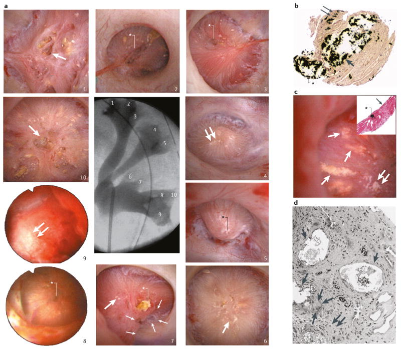
Figure 4. Renal papilla of a patient who formed brushite stones.
a | Endoscopic images of all ten papilla. The numbering corresponds to the x-ray image of the collection system. Papilla 1, 6, 7 and 10 show severe morphological changes including pitting (large arrows), flattening, plugs (black asterisk, papilla 7) and yellow plaque. Papilla 2, 3, 5 and 8 show small pits (white asterisks) whereas papilla 4, 7 and 9 show small sites of interstitial plaque (double arrows). b | Scattered inner medullary collecting ducts (IMCDs) and Bellini duct (BD) filled with yellow crystalline deposits that have dilated the tubular lumens (arrow) and protrude from the opening of the BD (asterisk). Small sites of interstitial plaque are also visible (double arrows). c | Sites of yellow plaque (arrows) in the lumens of IMCD (asterisk in insert) just beneath the urothelium (arrow in insert) and near sites of interstitial plaque (double arrows). d | showing extensive injury in the lining cells of IMCDs clogged with mineral deposits (arrows). Regions of extensive interstitial fibrosis surround the plugged IMCDs (double arrows) and entrapped and injured adjacent thin loops of Henle (asterisk). Giant cells (arrowheads) were occasionally found near damaged IMCDs.

