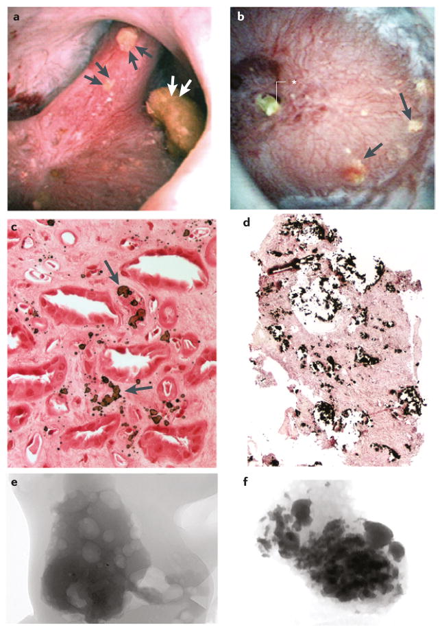
Figure 5. Renal papilla of patients who formed idiopathic apatite stones.
Patients who form apatite stones can be categorized into two groups on the basis of endoscopy images: those with a | normal appearing papillae and those with b | severe changes that resemble those of patients who form distal renal acidosis stones. Panel a | shows a papilla with three separate attached stones (double arrowheads) and no yellow plaque or Bellini duct plugs, whereas panel b | shows a papilla with numerous regions of yellow plaque (arrows) and a large BD plug (asterisk). Light microscopy images showing c | novel interstitial plaque structures (arrow) — a form of plaque that is unique to patients who form apatite stones — characterized by irregular, large and randomly distributed laminar structures of hydroxyapatite crystal and matrix, and d | numerous plugged inner medullary collecting ducts surrounding an area of extensive interstitial fibrosis in a papilla with severe changes. Micro-CT images of tubular deposits in biopsy samples from e | a normal appearing papilla with no deposits and f | a severely damaged papilla with numerous small deposits.

