Fig. 2.
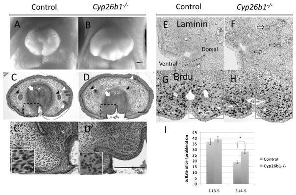
Abnormal cell proliferation and differentiation in the GTs of Cyp26b1−/− mutants. A and B: Frontal view of an E18.5 female GT in a control embryo and Cyp26b1−/− mutant. The size of the GT in the mutant is enlarged compared with that of the control. C, C′, D, and D′: Transverse section of the proximal GT in an E18.5 female; C′ and D′ are enlarged views of the framed area (dashed black line) in C and D. The development of the preputial gland (black arrowhead) and hair follicles (white arrowheads) under the genital skin is impaired in the Cyp26b1−/− mutants; mesenchymal condensation of the prospective corpus cavernosum penis is abnormal in the mutants (white arrow); ectopic blood vessels (area indicated by white frame) develop in the bilateral and dorsal mesenchyme of the UE in the mutants (C′ and D′); epithelium (black arrow) between the glans and prepuce is absent in the mutant (C, C′, D, and D′). E and F: Blood vessels indicated by Laminin immunostaining developed ectopically (hollow arrow) in the dorsal and bilateral mesenchyme of the UE at E14.5 in the mutant. To show the images of the mesenchyme on the dorsal side of the UE, the picture is displayed with an angled orientation, indicated by arrows. G and H: BrdU immunostaining of an E14.5 GT section. I: Cell proliferation assay. Framed area in G and H was counted, and the ratio of BrdU-positive cells was compared between the control and the mutant at E13.5 (n = 6) and E14.5 (n = 6). The asterisk (*) indicates significant statistical differences. All images of the cross-section are placed ventral side down unless otherwise indicated. Arrow with dashed stem in E indicates the ventral–dorsal direction of the GT. Scale bar, 200 μm. GT, genital tubercle; UE, urethral plate epithelium; BrdU, 5-BromodeoxyUridine.

