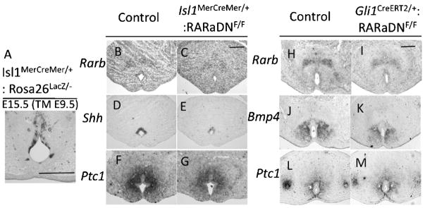
Fig. 5.
Impaired RA signaling reduces Shh and Bmp4 expression levels in the GT. A: The Isl1MerCreMer mouse line shows Tamoxifen (TM)-induced Cre expression in the UE. B and C: Rarb expression in the UE is reduced in the Isl1MerCreMer/+:RARaDNF/F mutant. D and E: Shh expression in the UE is reduced in the Isl1MerCreMer/+:RARaDNF/F mutant. F and G: Ptc1 expression in the bilateral mesenchyme of the UE is reduced in the Isl1MerCreMer/+:RARaDNF/F mutant. H and I: Rarb expression in the GT mesenchyme is reduced in the Gli1CreERT2/+:RARaDNF/F mutant. J and K: Bmp4 expression in bilateral mesenchyme of the UE is reduced in the Gli1CreERT2/+: RARaDNF/F mutant. L and M: Ptc1 expression in the bilateral mesenchyme of the UE is unaltered in the Gli1CreERT2/+: RARaDNF/F mutant. All images of the cross-section are placed ventral side down. Scale bar, 200 μm. GT, genital tubercle; RA, retinoic acid; UE, urethral plate epithelium; Bmp, bone morphogenetic protein.

