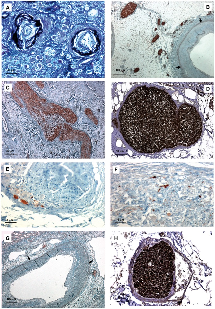
FIGURE 1.
Histologic features of native and transplanted kidneys in Patient 1. (A) Renal arterioles of native kidney featured concentric intimal thickening greater than thickness of media and concentric elastic duplication (score = 5; Weigert stain—bar = 40 μm). (B) In the native renal artery several nerves were observed within the adventitia, especially in the area close to the tunica media; nerve diameter ranged from 26 to 121 μm (NFP stain—bar = 100 μm). (C) Evidence of GAP43-positive nerve sprouting was noted in native ganglions close to arterial anastomoses (GAP43 stain—bar = 40 μm). (D–F) In renal nerves of native kidneys, TH-positive sympathetic efferent fibres (D) were more numerous than CGRP-positive sympathetic afferent fibres (E) and NOS-positive parasympathetic fibres (F) (D, bar = 10 μm; E, bar = 5 μm; F, bar = 5 μm). (G) Few GAP43-positive nerve axons could be observed in the renal artery from kidney transplant, distal to arterial anastomosis (GAP43 stain—bar = 100 μm). (H) In transplant renal artery, nerves consisted only of TH-positive sympathetic efferent fibres (TH stain—bar = 10 μm). (Adventitial–medial borders are highlighted by arrows.)

